|
Name of defect
|
Description
|
Method of control
|
Notes
|
|
Inclusion
|
a particle of alien material in the bulk of a crystal
|
1. Visual inspection under focused white light
2. Visual inspection through a microscope
|
Method 2 is used when extra high quality material is required
|
|
Block
|
a crystal part whose orientation in different from the orientation of the whole crystal
|
1. Visual inspection under focused white light
2. Visual inspection with a polariscope
|
|
|
Block mark
|
a boundary between parts of the crystal with different orientations
|
1. Visual inspection under focused white light.
2. Visual inspection with a polariscope
|
|
|
Twin
|
a crystal part whose lattice is a direct reflection of the lattice of the whole crystal
|
1. Visual inspection under focused white light.
2. Visual inspection with a polariscope
|
|
|
Slip line
|
a trace of plastic deformation of the crystal when atomic planes slip over each other. It looks like a straight line (stria) or several lines that can be revealed after polishing or annealing.
|
1. Visual inspection under focused white light.
2. Visual inspection with a polariscope
|
|
|
Bubble
|
a pore of vacuum space or gas in the bulk of a crystal which appears during crystallization
|
1. Visual inspection under focused white light.
2. Visual inspection with a polariscope.
3. Visual inspection through a microscope
|
Bubbles are always present is some II-VI crystals, e.g. ZnTe
Method 3 is used when extra high quality material is required
|
|
Microbubble
|
a bubble under 50 µm in diameter
|
1. Visual inspection under focused white light
2. Visual inspection with a polariscope
3. Visual inspection through a microscope
|
Bubbles are always present is some II-VI crystals, e.g. ZnTe
Method 3 is used when extra-high quality material is required
|
|
Macrobubble
|
a bubble over 50 µm in diameter
|
1. Visual inspection under focused white light;
2. Visual inspection with a polariscope
|
Bubbles are always present is some II-VI crystals, e.g. ZnTe
|
|
Bubble line
|
a gathering of bubbles extending along the crystallization front, the distance between the bubbles in less than 2 mm
|
1. Visual inspection under focused white light.
2. Visual inspection with a polariscope
3. Visual inspection through a microscope
|
Bubbles are always present is some II-VI crystals, e.g. ZnTe
Method 3 is used when extra-high quality material is required
|
|
Low grain boundary (lineage)
|
a boundary between parts of the crystal with slight orientation differences (up to 2 degrees)
|
1. Visual inspection with a polariscope
2. X-Ray diffraction
|
The defect is acceptable for LED and some optical applications
Method 2 is used for specific purposes
|
|
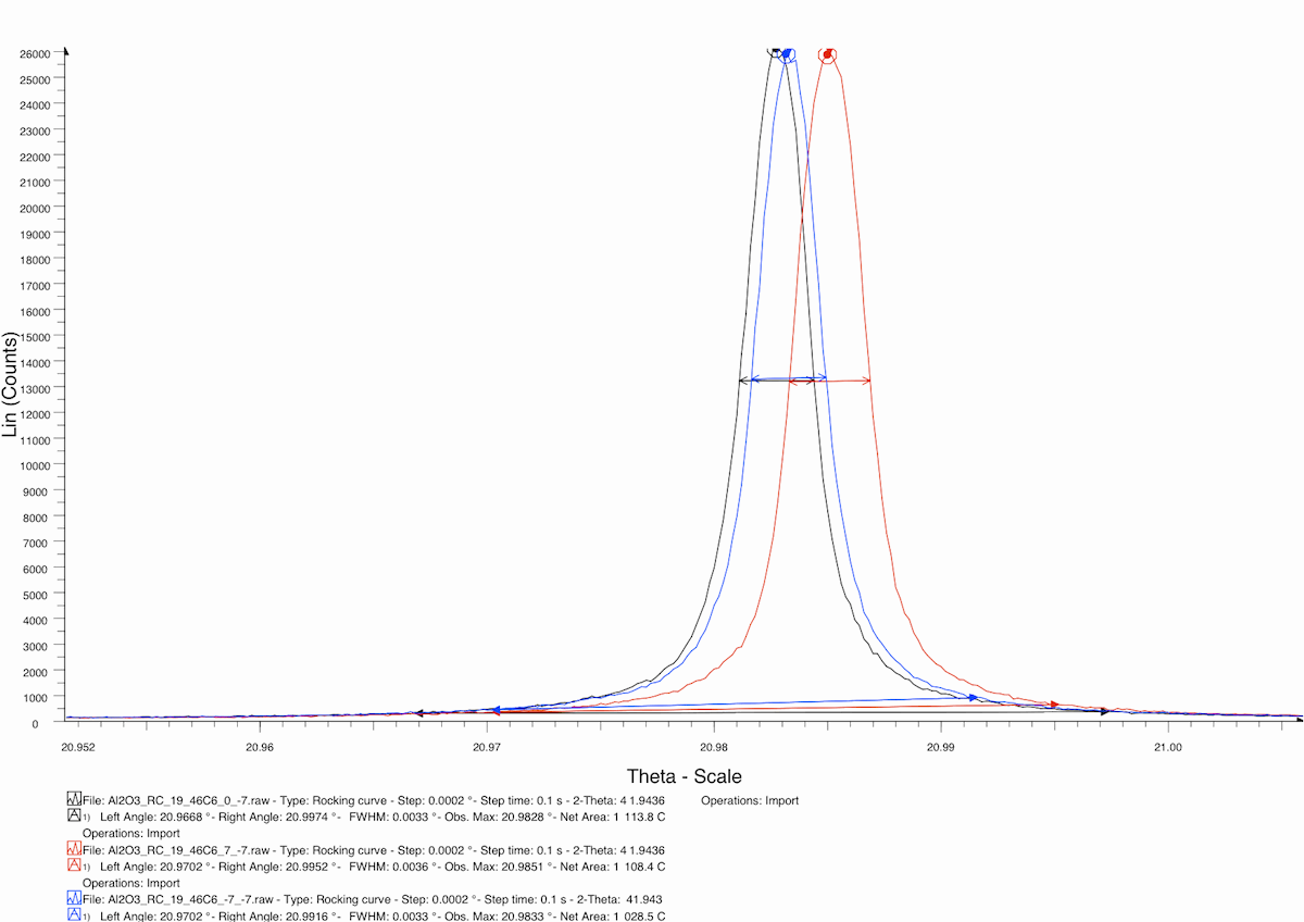
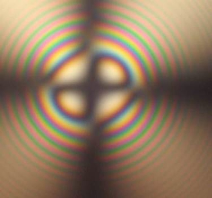
Soft low grain boundaries (soft lineage)
|
a boundary between parts of the crystal with slight orientation differences (a few minutes)
|
1. Visual inspection with a polariscope
2. Comparison of an interference picture of the tested crystal with an interference picture of an etalon crystal of the same thickness +/-10 mm and dislocation density of 5000 / cm2
3. X-Ray diffraction
|
The defect is acceptable for LED, RF and most optical applications.
Method 3 is used for specific purposes
|


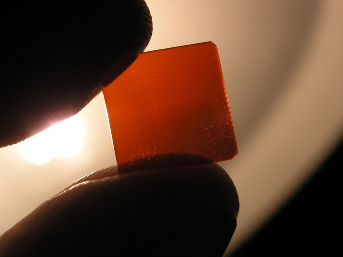
 Soft lineage in sapphire
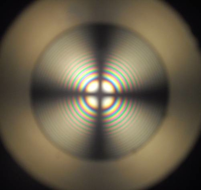 Etalon sapphire crystal